- 移动端


翌圣生物科技(上海)股份有限公司品牌商
19 年
手机商铺
商家活跃:
产品热度:
- NaN
- 0.7999999999999998
- 1.7999999999999998
- 0.7999999999999998
- 3.8
自营
翌圣生物(Yeasen)
试剂/耗材
已认证品牌介绍
翌圣生物科技(上海)股份有限公司是一家聚焦生命科学产业链上游核心原料,从事分子、蛋白和细胞三大品类生物试剂的研发、生产与销售的高新技术企业。核心产品覆盖qPCR系列、NGS系列、逆转录系列、核酸提取与纯化系列、PCR系列、分子克隆系列、体外转录系列、抗体、蛋白纯化系列、蛋白分析系列、重组蛋白、细胞分析系列、细胞培养系列、细胞转染系列、报告基因检测系列等多个品类,广泛应用于生命科学研究、诊断检测和生物医药等领域。
代理
Sino Biological
抗体/蛋白质/抗原/多肽
已认证品牌介绍
义翘神州(Sino Biological)始终秉承“为全球生命科学研究人员提供高品质的重组蛋白和抗体等科研工具试剂以及一站式技术服务”的理念,坚持自主开发,不断提升技术水平,以创新的精神、先进的技术、严格的质量控制体系为基石,全面致力于以客户需求为导向,不断优化产品品质和服务体验,打造国际一流生物试剂品牌,赢得全球客户的信赖和好评。
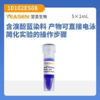
Annexin V-Alexa Fluor 488/7-AAD细胞凋亡检测试剂盒
¥388
品牌商
翌圣生物科技(上海)股份有限公司
入驻年限:19 年
- 联系人:
翌圣生物
- 所在地区:
上海
- 业务范围:
体外诊断、耗材、技术服务、抗体、ELISA 试剂盒、细胞库 / 细胞培养、试剂
- 经营模式:
生产厂商
推荐产品

公司新闻/正文
Annexin-V-Alexa 568 细胞凋亡及细胞死亡
2338 人阅读发布时间:2011-10-17 23:37
Annexin-V-Alexa 568,现货供应!Roche经典产品!
Application
The analysis of phosphatidylserine on the outer leaflet of apoptotic cell membranes is performed using Annexin-V-Alexa 568 in combination with a DNA stain (e.g., BOBO-1) to differentiate apoptotic cells from necrotic cells. Apoptotic and necrotic cells can also be differentiated by cell surface markers.
Benefits
- Detection flexibility: Analysis of results with flow cytometry and/or fluorescence microscopy
Alexa is a red dye, which can be used for double or triple staining with other fluorescent markers (e.g., fluorescein-labeled surface markers) - Accuracy: Accurately distinguish apoptotic cells from necrotic cells
- Stability of results: High photostability (long observation possible)
- Rapid and simple procedure
Product Description
Alexa 568 is a fluorescent label. The Annexin-V-Alexa 568 conjugate can be easily analyzed on a 488-nm laser Fluorescent Analytical Cell Sorter (FACS) machine, or using a fluorescence microscope. Excitation is in the range of 488 - 596 nm with emission above 600 nm.
Additional secondary labeling is also possible. For example, membrane surfaces can be stained with fluorescein-labeled monoclonal antibodies for further cellular characterization.
Additional secondary labeling is also possible. For example, membrane surfaces can be stained with fluorescein-labeled monoclonal antibodies for further cellular characterization.
Background Information
In the early stages of apoptosis, changes occur on the cell surface. One of these plasma membrane alterations is the translocation of phosphatidylserine (PS) from the inner side of the plasma membrane to the outer layer, thereby exposing PS at the external surface of the cell. Fadok et al. showed that macrophages specifically recognize PS exposed on the surface of lymphocytes during apoptosis. This recognition and subsequent phagocytosis of apoptotic cells and bodies protects organisms from exposure to cellular compounds that lead to inflammation and usually accompany necrosis.
Annexin-V is a Ca2+-dependent, phospholipid-binding protein with a high affinity for PS. This protein can therefore be used as a sensitive probe for PS exposure on the outer leaflet of the cell membrane, and is suited to detect apoptotic cells. Since necrotic cells also expose PS as a result of lost membrane integrity, apoptotic cells must be differentiated from these necrotic cells. The simultaneous application of a DNA stain (used for dye-exclusion tests [e.g., BOBO-1]) allows the discrimination of necrotic cells from the Annexin-V positively stained cell cluster.
Contents
500 µl, ready-to-use solution, for 250 tests
Typical Experiment
Figure 1: Discrimination between apoptotic and necrotic U937 cells treated with camptothecin (CAM) and stained with Annexin-V-Alexa 568 (red) and BOBO-1 (green).
我公司所售全部产品均为科研用途!
免费电话:4006-111-883
免费电话:4006-111-883
| 上海前尘生物科技有限公司 | |
| 公司地址: | 上海市徐汇区南丹东路18号12楼A座 |
| 联系电话: | 021-34615995 021-34615975 |
| 传真: | 021-34615995-8008 021-34615975-8008 |
| 移动电话: | 13918775203 (周末和紧急情况下联系) |
| E-mail: | sales@qcbio.com info@qcbio.com |
| 北京分公司 | |
| 公司地址: | 北京朝阳区大屯路2号南沙滩35号楼科华商务大厦613室 |
| 联系电话: | 010-64847623 010-64855926 |
| 传真: | 010-64847623-8008 010-64855926-8008 |
| 移动电话: | 18910229156 (周末和紧急情况下联系) |
| E-mail: | lwh@qcbio.com |
| 南京办事处 | |
| 公司地址: | 南京市鼓楼区中央路323号利奥大厦1402室(和新模范马路路口交叉) |
| 联系电话: | 025-83535567 |
| 传真: | 025-83535565 |
| 移动电话: | 18061695143(周末和紧急情况下联系) |
| Email: | qcbio.wu@gmail.com |